
- Sudden, severe, electric-shock-like pain on one side of the face
- Diagnosis is made chiefly from a careful history — expertise matters
- Most patients respond initially to medication (carbamazepine)
- When medication fails, surgery offers excellent, lasting relief
- Mr Russell manages 100+ new patients a year and offers every treatment option
- Brief episodes of pain lasting from less than a second to a couple of minutes.
Often the attacks come in waves, with episodes lasting days to weeks then settling down for a period before returning. - Pain is usually stabbing, shooting, sharp or “like electricity”.
- Pain is triggered by light facial touch such as talking, eating, brushing teeth, wind, etc
- Pain is located in the face, most often the cheek, lips or jaw, but may also involve the side of the nose, inside of the mouth, eye or forehead.
- Pain is usually confined to one side of the face, but may very occasionally affect both sides.
- Pain usually initially responds to carbamazepine (Tegretol) or oxcarbazepine (Trileptal).
Internationally, trigeminal neuralgia is now classified as classical (due to compression of the nerve by a blood vessel), secondary (due to another condition such as multiple sclerosis or a tumour), or idiopathic (no cause found).4Each type may be either purely paroxysmal, with brief attacks only, or accompanied by concomitant continuous pain — a constant background aching or burning between attacks, which occurs in roughly a quarter to a half of patients.4 A more detailed surgical classification (the Burchiel system) is also used:5
- Trigeminal neuralgia type 1 (TN1): classical form where episodic lancinating pain predominates, also known as “typical TN”.
- Trigeminal neuralgia type 2 (TN2): atypical form where pain is aching, throbbing or burning and constant for >50% of the time, also known as “atypical TN”. TN2 is further divided into TN2a, where the constant pain has evolved from a preceding history of TN1, and TN2b, where the constant pain has been present from the onset without an earlier episodic phase.6
- Trigeminal neuropathic pain (TNP): pain related to unintentional injury to the nerve from facial trauma, ENT surgery, oral surgery, posterior fossa surgery or stroke.
- Trigeminal deafferentation pain (TDP): pain in an area of facial numbness, related to intentional injury to the nerve such as rhizotomy, neurectomy, gangliolysis or other denervating procedure.
- Symptomatic trigeminal neuralgia (STN): pain associated with multiple sclerosis.
- Persistent idiopathic facial pain (PIFP), previously called atypical facial pain: a separate, poorly understood chronic facial pain that does not follow the trigeminal neuralgia pattern and is distinguished from it.
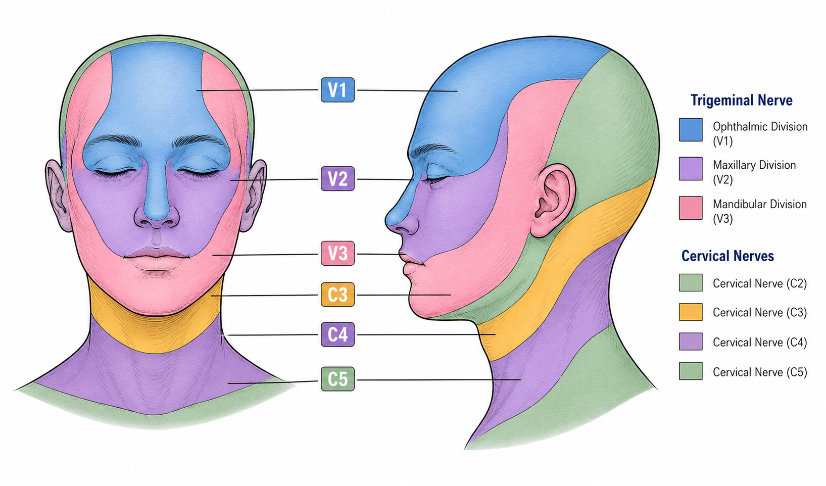
The three divisions of the trigeminal nerve — ophthalmic (V1), maxillary (V2) and mandibular (V3).
Ordinary pain-relief medications such as paracetamol, codeine and other opioids do not generally work well for this condition. The medications that do help are typically anti-seizure (anticonvulsant) drugs — though it is worth stressing that trigeminal neuralgia is neither a seizure nor a form of epilepsy; these drugs work by calming the abnormal nerve signalling that produces the pain.
Carbamazepine (Tegretol) is the first-line treatment and has the strongest evidence of any drug for trigeminal neuralgia, with the great majority of patients gaining good relief when it is first started.8,9 It is begun at a low dose and increased gradually until the pain settles. Common side effects include drowsiness, dizziness, unsteadiness and nausea, and less commonly a low blood-sodium level or a skin rash; rare but serious reactions affecting the blood cells, liver or skin can occur, so occasional blood tests are recommended. In people of Asian descent a blood test (HLA-B*1502) is advised beforehand to identify the small group at higher risk of a severe skin reaction.
Oxcarbazepine (Trileptal) is a closely related first-line alternative of similar effectiveness that is often better tolerated and has fewer drug interactions, though it is somewhat more likely to lower the blood-sodium level, which is checked with blood tests.8 It is, however, considerably more expensive than carbamazepine.
If the first-line drugs fail or are not tolerated, a number of second-line agents may be used, either on their own or added to a first-line drug — these include gabapentin (Neurontin), sodium valproate, pregabalin (Lyrica), lamotrigine (Lamictal) and baclofen.8
In a severe, resistant crisis — an acute exacerbation with uncontrollable pain — hospital admission may be required, and intravenous medications such as lidocaine, phenytoin (or fosphenytoin) and ketamine can be used to bring the pain under control.8
- Microvascular decompression (MVD)
- Percutaneous needle rhizotomy (PNR)
- Stereotactic radiosurgery (SRS)
Microvascular decompression (MVD) is the only treatment that directly addresses the underlying cause of trigeminal neuralgia — the blood vessel compressing the trigeminal nerve where it leaves the brainstem. It offers the greatest chance of long-lasting, medication-free relief and is the preferred surgical option for patients with neurovascular compression.
The operation is performed under general anaesthetic. A small opening is made in the skull behind the ear (a retrosigmoid craniotomy) and, using a surgical microscope, the trigeminal nerve is identified at the brainstem. The offending artery is gently moved away from the nerve and held in its new position with a small piece of Teflon and tissue glue — a technique known as transposition. Only rarely is a vein responsible; it is almost always an artery.
An MVD offers an approximately 95% chance of immediate and complete resolution of the pain, with the ability to come off all medication. Most recurrences, when they occur, happen within the first two years, after which the yearly risk becomes very low — under 1% per year. In large published series, around 70% of patients remained free of pain without any medication ten years after surgery.10
The likelihood of any significant complication is very low. The main risks are:
- Double vision — an approximately 5% risk of temporary (and less than 1% permanent) weakness of the fourth (trochlear) nerve, causing double vision that is particularly noticeable when reading.
- Facial numbness — a 1–2% chance.
- Facial weakness — a less than 1% chance.
- Reduced hearing on the operated side — a 1–3% chance.
- Brainstem injury — a less than 1 in 1000 chance, but potentially serious, with severe neurological consequences such as paralysis, sensory disturbance, or difficulty swallowing or speaking.
- Return to theatre for bleeding — a less than 1% chance.
- Infection — a 1–2% chance, often delayed by several months.
- Cerebrospinal fluid (CSF) leak — a 1–2% chance.
- General anaesthetic risks — including clots in the legs or lungs (DVT/PE), heart attack, stroke, severe allergic reaction. Taking the operation as a whole, the risk of death is very low — in the order of 0.2% (about 1 in 500).
Patients spend the first night in the Intensive Care Unit (ICU) prior to being transferred to the ward. Typical inpatient stay is between four and six nights. Significant headache, nausea and vomiting are common in the first day or two though quickly settle. Someone will need to care for you at home for the first week after discharge.
You will need to review with your GP 7–10 days after surgery to check the wound and confirm your recovery is progressing well. Sutures are self-dissolving and should not be removed. Providing the trigeminal pain has resolved, begin weaning trigeminal medication after 24 hours. Most patients are able to stop all trigeminal medication within a few weeks. You will review with the surgeon six weeks after surgery. Most people resume normal daily activities within one to two weeks, although usually unable to work for at least six weeks, and sometimes longer.
This is a day procedure and usually the patient goes home later that day or the following morning. It is less invasive than an MVD but more than SRS. It is typically performed under general anaesthesia, although it can also be done under sedation. A specially designed needle is guided using X-ray and CT navigation to where the trigeminal nerve exits the skull. The nerve is treated with a chemical (glycerol), heat (radiofrequency electrocoagulation, RFE) or a balloon compression. All of these procedures work by deliberately damaging the nerve — in essence, trading the pain for permanent facial numbness. As a rule, the greater the numbness produced, the longer the period of freedom from pain. Treatment is therefore usually begun with glycerol, which creates the least numbness and allows a person to become accustomed to a numb face before progressing, if needed, to techniques that produce greater numbness. Most patients (around 75–80%) gain significant or complete relief of their pain. This relief typically lasts between one and four years — sometimes shorter, sometimes longer — after which the procedure can be repeated.11
Facial numbness is the major drawback to a rhizotomy, and some reduction in facial sensation is almost inevitable. Although unsettling at first, once a person has become accustomed to it — usually within one to two months — it is not normally too troubling. Other risks include weakness of chewing on the treated side (around 5%), double vision (less than 1%), a less than 1% risk of injury to the nearby carotid artery (which can rarely cause a stroke or an abnormal artery-to-vein connection needing further treatment), meningitis (less than 1%), the rare possibility of anaesthesia dolorosa (constant, difficult-to-treat pain despite a numb face), and the usual anaesthetic risks — clots in the legs or lungs (DVT/PE), heart attack, stroke, severe allergic reaction, and a remote risk of death.
Percutaneous needle rhizotomy is generally reserved for:
- atypical trigeminal neuralgia
- trigeminal neuralgia secondary to multiple sclerosis
- patients unfit for an MVD
- patients not wishing to undergo an MVD
- Katusic S, Beard CM, Bergstralh EJ, Kurland LT. Incidence and clinical features of trigeminal neuralgia, Rochester, Minnesota, 1945-1984. Ann Neurol. 1990;27(1):89-95. doi:10.1002/ana.410270114
- De Toledo IP, Conti Réus J, Fernandes M, et al. Prevalence of trigeminal neuralgia: a systematic review. J Am Dent Assoc. 2016;147(7):570-576. doi:10.1016/j.adaj.2016.02.014
- Trigeminal Neuralgia Association Australia. Awareness for trigeminal neuralgia. Accessed July 2026. https://tnaaustralia.org.au/awareness
- Cruccu G, Finnerup NB, Jensen TS, et al. Trigeminal neuralgia: new classification and diagnostic grading for practice and research. Neurology. 2016;87(2):220-228. doi:10.1212/WNL.0000000000002840
- Burchiel KJ. A new classification for facial pain. Neurosurgery. 2003;53(5):1164-1167. doi:10.1227/01.neu.0000088806.11659.d8
- Eller JL, Raslan AM, Burchiel KJ. Trigeminal neuralgia: definition and classification. Neurosurg Focus. 2005;18(5):E3. doi:10.3171/foc.2005.18.5.4
- Bendtsen L, Zakrzewska JM, Heinskou TB, et al. Advances in diagnosis, classification, pathophysiology, and management of trigeminal neuralgia. Lancet Neurol. 2020;19(9):784-796. doi:10.1016/S1474-4422(20)30233-7
- Bendtsen L, Zakrzewska JM, Abbott J, et al. European Academy of Neurology guideline on trigeminal neuralgia. Eur J Neurol. 2019;26(6):831-849. doi:10.1111/ene.13950
- Gronseth G, Cruccu G, Alksne J, et al. Practice parameter: the diagnostic evaluation and treatment of trigeminal neuralgia. Neurology. 2008;71(15):1183-1190. doi:10.1212/01.wnl.0000326598.83183.04
- Barker FG 2nd, Jannetta PJ, Bissonette DJ, Larkins MV, Jho HD. The long-term outcome of microvascular decompression for trigeminal neuralgia. N Engl J Med. 1996;334(17):1077-1083. doi:10.1056/NEJM199604253341701
- Lopez BC, Hamlyn PJ, Zakrzewska JM. Systematic review of ablative neurosurgical techniques for the treatment of trigeminal neuralgia. Neurosurgery. 2004;54(4):973-982. doi:10.1227/01.neu.0000114867.98896.f0
- Tuleasca C, Régis J, Sahgal A, et al. Stereotactic radiosurgery for trigeminal neuralgia: a systematic review: International Stereotactic Radiosurgery Society practice guidelines. J Neurosurg. 2018;130(3):733-757. doi:10.3171/2017.9.JNS17545