
- Shooting or stabbing pain over the back and top of the scalp
- Arises from the greater, lesser and third occipital nerves
- Estimated incidence around 3.2 per 100,000 per year
- Diagnosis is clinical; a nerve block can help confirm it
- Treated with medication, nerve blocks and, in selected cases, surgery
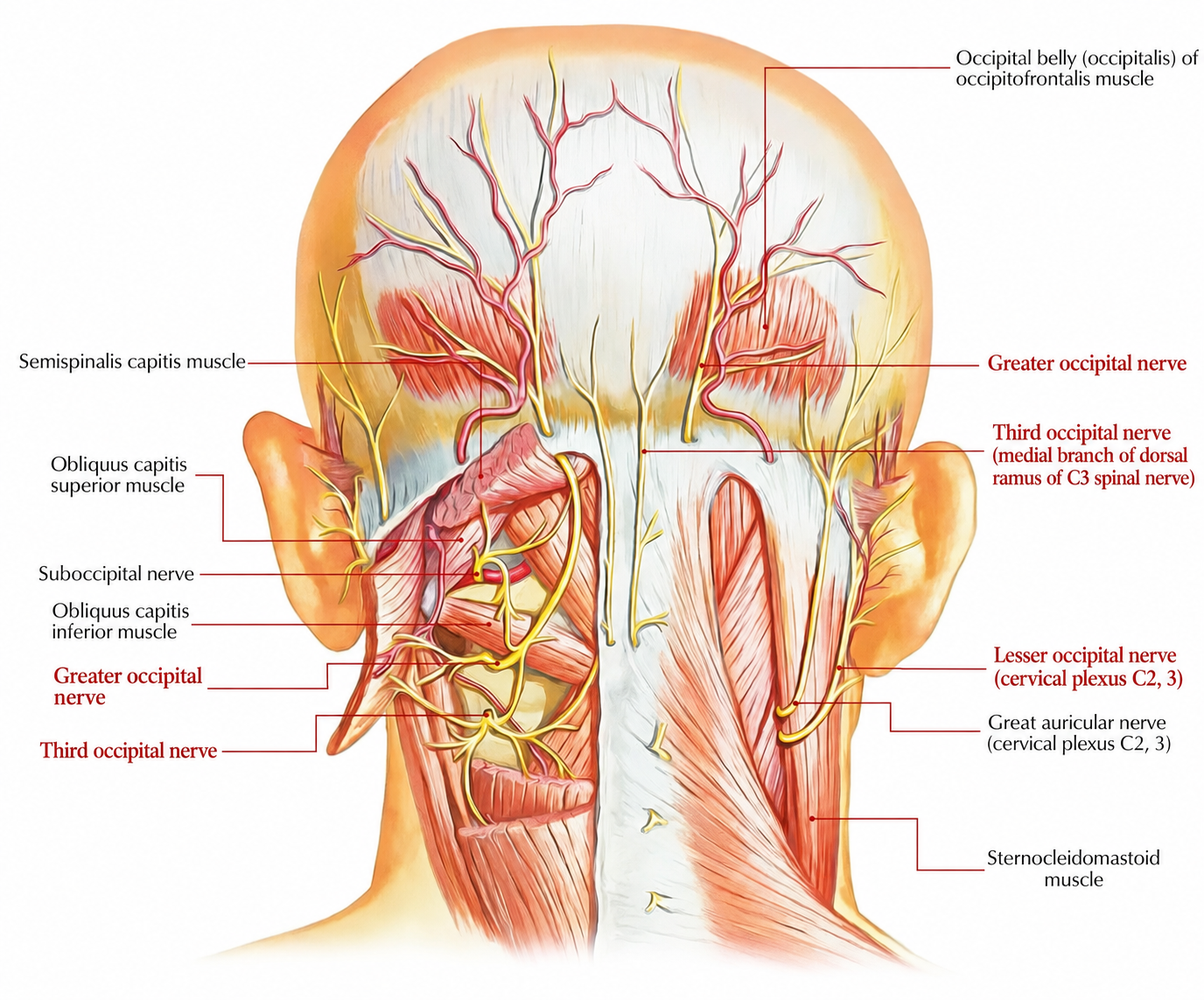
Figure 1. Diagrammatic representation of the occipital nerves (greater, lesser and third) and related structures of the posterior scalp and neck.
The occipital nerves arise from the upper cervical nerve roots (primarily C2 and C3) and travel upwards through muscles and connective tissue at the back of the neck before piercing through the muscles to supply the scalp. Irritation, compression, or injury at any point along this course can cause ON. In the majority of patients no specific structural cause is found — the pain is either idiopathic or attributed to irritation or entrapment of the nerves as they pass through the neck muscles. When an identifiable cause is present, it is most often one of the following:
- Nerve entrapment (the most common mechanism) — the GON pierces through the semispinalis capitis muscle in approximately 90% of people and through the trapezius aponeurosis in approximately 45%, creating potential anatomical sites where the nerve may become compressed or entrapped 2
- Trauma — whiplash injury, direct impact to the back of the head or neck, or prior cervical surgery causing scarring around the nerve
- Degenerative cervical spine disease — arthritis or disc disease of the upper cervical spine (C1–C2, C2–C3) compressing nerve roots
- Less common causes — tumours or other mass lesions, inflammatory conditions such as rheumatoid arthritis of the upper cervical joints, and rarely, giant cell arteritis
The International Headache Society (ICHD-3) defines ON as unilateral or bilateral paroxysmal, shooting or stabbing pain in the distribution of the greater, lesser, or third occipital nerves, sometimes accompanied by reduced sensation or altered sensation (dysaesthesia) in the affected area, and commonly associated with tenderness over the involved nerve(s). 3
In practice, symptoms typically include:
- Sudden, sharp, shooting or electric shock-like pain starting at the base of the skull and radiating upward over the scalp toward the forehead and behind the eye
- Tenderness on firm pressure over the greater occipital nerve (at the back of the skull, roughly one third of the way between the mastoid and the midline) — this is a characteristic and important examination finding
- Scalp sensitivity (allodynia) — the scalp feels painful or abnormally tender to light touch, making it difficult to lie on a pillow, brush hair, or wear glasses or a hat
- Pain that may radiate forward to the temple, forehead, or behind the eye on the affected side
The pain is typically unilateral, affecting one side in around 80% of people, 4 and usually begins on one side; it is only rarely bilateral at onset, though it may extend to both sides in the remainder. It may occur in bursts or persist as a more constant ache with intermittent sharp exacerbations.
ON is one of the most commonly misdiagnosed conditions in headache medicine. 3 Conditions frequently confused with ON include:
- Migraine — the most common diagnostic overlap; migraine is a primary brain-generated headache, typically throbbing, often with nausea, and associated with light and sound sensitivity; it lacks the focal nerve tenderness characteristic of ON. Importantly, occipital nerve blocks can relieve both ON and migraine, making the block non-specific as a diagnostic test 5
- Cervicogenic headache — referred pain from the upper cervical joints or discs, typically provoked by neck movement, side-locked, and non-lancinating; does not have the paroxysmal electric-shock quality of ON
- Tension-type headache — dull, bilateral, band-like pressure, without the nerve-distribution pattern or focal tenderness of ON
- Cluster headache — severe periorbital and temporal pain occurring in cyclical patterns, associated with autonomic features (tearing, nasal congestion, drooping eyelid)
- C2 radiculopathy — pain from C2 nerve root compression in the cervical spine; distinguished by cervical imaging and examination findings
- Greater occipital neuritis from giant cell arteritis — inflammation of the scalp arteries in older patients; distinguished by elevated inflammatory markers (ESR, CRP), jaw claudication, and scalp artery tenderness
- Post-herpetic neuralgia — persistent burning pain following shingles (herpes zoster) in the occipital distribution; distinguished by history of shingles rash
Simple measures that are often helpful as initial management include:
- Local heat application to the back of the neck
- Gentle massage and stretching of the suboccipital muscles
- Rest and activity modification to avoid neck postures that aggravate pain
- Non-steroidal anti-inflammatory drugs (NSAIDs) and muscle relaxants may provide modest short-term relief, though they are generally insufficient as sole long-term treatment
Medications used for ON are the same as those used for other neuropathic pain conditions, with carbamazepine being the traditional first-line agent. It is important to note that no large randomised controlled trials have been performed specifically in ON, and medical treatment recommendations are largely extrapolated from evidence in other neuralgias and neuropathic pain conditions. 6
Carbamazepine (Tegretol) is the most established first-line oral medication, widely used for neuralgias. It stabilises abnormally excitable nerve membranes. It is started at a low dose — typically 100–200 mg/day, with the lower dose used in smaller or elderly patients — and increased gradually. Common side effects include drowsiness and dizziness; blood monitoring for sodium levels and blood count is required. 5
Pregabalin (Lyrica) and Gabapentin (Neurontin) are anticonvulsant medications with established evidence in neuropathic pain. Gabapentin 300–600 mg at bedtime is a commonly used alternative. 5
Amitriptyline (a tricyclic antidepressant) is a useful second-line agent, particularly when sleep disruption accompanies pain. 5
Oxcarbazepine (Trileptal) is a carbamazepine analogue that is often better tolerated, with fewer drug interactions.
Other agents that may be helpful include baclofen, lamotrigine, nortriptyline 30–50 mg at bedtime, and topical agents. 5
Standard over-the-counter pain relievers such as paracetamol and ibuprofen are generally insufficient as primary treatment for ON.
For patients whose pain returns between nerve blocks or who require frequent repeat injections, several more durable minimally invasive options are available:
Pulsed radiofrequency (PRF) treatment — delivers pulsed electrical energy around the occipital nerve or C2 dorsal root ganglion (DRG), reducing pain signalling without destroying the nerve. A clinical study of 10 patients found PRF to provide significant pain relief with a good safety profile. 7
Radiofrequency ablation (RFA) — delivers controlled heat to the C2 dorsal root ganglion or third occipital nerve, producing a longer-lasting reduction in pain transmission. In a study of 40 patients with cervicogenic headache or occipital neuralgia, RFA of the C2 dorsal root ganglion and/or third occipital nerve produced at least 80% pain relief in around 70% of appropriately selected patients. 8
Occipital nerve decompression (neurolysis)
Occipital nerve removal (neurectomy)
- Koopman JSHA, Dieleman JP, Huygen FJ, de Mos M, Martin CGM, Sturkenboom MCJM. Incidence of facial pain in the general population. Pain. 2009;147(1-3):122-127. doi:10.1016/j.pain.2009.08.023
- Bovim G, Bonamico L, Fredriksen TA, Lindboe CF, Stolt-Nielsen A, Sjaastad O. Topographic variations in the peripheral course of the greater occipital nerve: autopsy study with clinical correlations. Spine (Phila Pa 1976). 1991;16(4):475-478. doi:10.1097/00007632-199104000-00017
- Djavaherian DM, Guthmiller KB. Occipital neuralgia. In: StatPearls. Treasure Island, FL: StatPearls Publishing; 2024. https://www.ncbi.nlm.nih.gov/books/NBK538281
- Melchior AG, Al-Khazali S, Christensen RH, Al-Khazali HM, Ashina H. Epidemiology and clinical features of occipital neuralgia: a systematic review and meta-analysis. Cephalalgia. 2025;45(2):3331024251317595. doi:10.1177/03331024251317595
- Barmherzig R, Kingston W. Occipital neuralgia and cervicogenic headache: diagnosis and management. Curr Neurol Neurosci Rep. 2019;19(5):20. doi:10.1007/s11910-019-0937-8
- Choi I, Jeon SR. Neuralgias of the head: occipital neuralgia. J Korean Med Sci. 2016;31(4):479-488. doi:10.3346/jkms.2016.31.4.479
- Choi HJ, Oh IH, Choi SK, Lim YJ. Clinical outcomes of pulsed radiofrequency neuromodulation for the treatment of occipital neuralgia. J Korean Neurosurg Soc. 2012;51(5):281-285. doi:10.3340/jkns.2012.51.5.281
- Hamer JF, Purath TA. Response of cervicogenic headaches and occipital neuralgia to radiofrequency ablation of the C2 dorsal root ganglion and/or third occipital nerve. Headache. 2014;54(3):500-510. doi:10.1111/head.12295
- Staudt MD, Hayek SM, Rosenow JM, et al. Congress of Neurological Surgeons systematic review and evidence-based guidelines for occipital nerve stimulation for the treatment of patients with medically refractory occipital neuralgia: update. Neurosurgery. 2023;93(3):493-495. doi:10.1227/neu.0000000000002578
- Li F, Ma Y, Zou J, et al. Micro-surgical decompression for greater occipital neuralgia. Turk Neurosurg. 2012;22(4):427-429. doi:10.5137/1019-5149.JTN.5234-11.1
- Ducic I, Felder JM 3rd, Khan N, Youn S. Greater occipital nerve excision for occipital neuralgia refractory to nerve decompression. Ann Plast Surg. 2014;72(2):184-187. doi:10.1097/SAP.0b013e3182898736